Friday, October 10, 2014

First "Second Opinion"

Thanks again to AVMsurvivors forum, I learnt about Barrow Neurological Institute (BNI) and their Second Opinion Program.

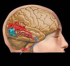
BNI is located in Phoenix, AZ and is a world renowned neurosurgical center.  In fact, it is the world's largest neurological disease treatment center with 11 dedicated Neurosurgical Operating Rooms performing about 100 surgeries a week. Yes, 100 brain surgeries a week on patients from all over the world. To top it of, AVM's grading scale, called Spetzler-Martin scale is actually named after BNI's Director Dr. Robert Spetzler.

So when it comes to AVMs, BNI's opinions matter a lot and I was pleasantly surprised to find out I can have all that world class consultation for just $100 via their remote second opinion program. All I had to do was fill out a short form online and upload my MRI images.

I immediately signed up and uploaded my MRI images. Within a few hours, someone reached out and asked for my MRI report and Visual Field Test report. I scanned and emailed them right away and waited to hear back.

In less than 24 hours, I had my report in my inbox. It was short, but not sweet. Here it is:

"I would recommend treatment given the fixed neurological deficit (left quadrantinopsia). The optimal treatment would be preoperative embolization (which I would do) followed by surgical resection (by Dr. Spetzler). If you would like to be seen in consultation and have your surgery at Barrow Neurological Institute, please contact Stephanie at 602-406-xxxx to inquire about arranging an appointment. We would be delighted to be of help in any way possible.

Sincerely, 
Felipe C. Albuquerque, MD"

So, their choice of treatment is exactly the same as the one outlined by my local neuro - embolization followed by craniotomy. But she was against the treatment and BNI is recommending it.

I thought about it for a night and realized I was not ready to give in either way yet. So, I sent them an email about my local neuro's recommendation against any treatment and asked BNI if they wanted to review my angiogram images and make another recommendation. Instead of asking for my angiogram images, I got this email back form BNI:

"The surgeon that reviewed feels that risk of surgery is minimal compared to the risk of a rupture. He feels you should have embolization and then surgery. The doctor used the imaging you had prior and has recommended surgery. If you are interested in coming for treatment, please let me know."

So, all I have now are 2 very conflicting recommendations to chose from. Confused, I again sought help at the AVMsurvivors forum and posted my predicament there. Got several responses in a short period.

  • One was skeptical about the local surgeon's recommendation to not treat an "operable" AVM
  • A few simply asked me to put my faith on BNI's experience and reputation
  • And others suggested getting another opinion (apparently this is very common)
Given the high risk either way, I have decided to seek another opinion to break the tie. But I want to do this in person and at a very reputable institute.

After a quick research, I zeroed in on Johns Hopkins Hospital in Baltimore MD. I chose this for some obvious reasons
  • It had been ranked No. 1 hospital in US for 21 consecutive years by US News
  • Its Neurosurgical practice has been ranked consistently among the top 3 in US and
  • More importantly, it is drivable from my home in Richmond, VA
And a (doctor) friend who used to work at Johns Hopkins recommended I consult with Dr. Rafael Tamargo. I looked him up and quickly realized he is one of the premier neurosurgeons in the whole world, especially in Cerebrovascular Neurosurgery. 

So, Dr. Tamargo and Johns Hopkins it is. I will leave my AVM alone (and hope it will leave me alone) or get it treated based on his recommendation. But the earliest appointment I could get was for mid November. Too bad, patience is not one of my virtues.


But I have something else to look forward to next week. I am flying back home for a week and I will finally let my wife know about this mess. So far, only one friend and one of my brothers know about this. I desperately wanted to tell my wife several times (especially when I went thro' the cerebral angiogram procedure all by myself), but I just couldn't do it over the phone. So there is going to be some drama next week :)

Tuesday, October 7, 2014

Maintain the Status Quo?

I visited my neurosurgeon this afternoon with the report and images from my angiogram. She said she had already reviewed them with the Interventional Radiologist and gave me the rundown.

Here is an actual image of my AVM from the angiogram images. The dark patch in the back of my brain is the AVM cluster (nidus).


First, the bad news:


  • My AVM nidus (or the AVM nest) was actually larger than the MRIs had indicated - 5.9cm x 4 cm x 3.9 cm
  • Remember I had mentioned in the last post about how the nidus was spread around a bit instead of being in a nice little clump? It turns out there is actually a term for it - Diffuse AVM. And mine was diffused quite a bit in my Occipital lobe.
  • She said it is at least a Grade 4 AVM.
  • Given its size and its diffused nature, she immediately ruled out radiation (Gamma Knife) as a treatment option.
  • Again, given its size and diffused nature, she said the treatment, if it were to be done, will involve 3 or 4 separate embolizations (spread over as many months) and followed finally by a craniotomy
  • And the craniotomy itself will be major surgery lasting several hours 
  • And finally, with the treatment, there is 80% chance of me losing good portion of my left side vision
At this point, I was ready to stick my fingers into my ears and sing something loud.

Now, the good news:

  • The speed of the blood flowing through the nidus is relatively slow, so the risk of bleeding is a bit lower than usual. 
  • Given how messed up my nidus is and all the risks associated with every step of the treatment (each embolization carries the risk of hemorrhaging, vision loss, etc.), she recommended that I leave the AVM alone and not treat it at all. Do nothing? I can do that. 
In short, the risks associated with the treatment outweigh the benefits, so it is better to take my chances (and push my luck) with an AVM rupture in the future.

My feelings were mixed at this point. I was frustrated a bit since I couldn't get it treated,  I was relieved a bit since I didn't have to get it treated, and I was scared a bit since I will have to live with this in my brain for the rest of my life and it could bleed anytime.

And then she said something that, I felt, pushed me back to square one - 
"My recommendation is to do nothing, but you will find a lot of 'trigger happy' neurosurgeons who will encourage you get this treated soon. And don't think I am afraid to treat this, I just think it is better if you leave it alone"

Sigh. Thats when I realized she never used the word "inoperable". But I was glad that she was being honest and open about this. She advised me to get a second opinion and recommended another neurosurgeon (her mentor) in another Swedish hospital in Seattle.

So, now I will have to get another opinion (or two). But I am going to cast a wider net across the US and look for hospitals and doctors who specialize in treating complex AVMs.


Wednesday, October 1, 2014

Cerebral Angiogram

I went to bed around 11:30 PM last night, but woke up in a few hours around 3 AM. I was anxious and restless and my angio was scheduled for 7:30 AM. I think I slept for a couple more hours between 4 and 6.

I reached the Radiology Dept. at Swedish Hospital around 7:15 AM and filled out a couple of forms, and then a nurse took me inside. I changed to a hospital gown and laid down on a bed. After the usual things a nurse does, an IV line was placed in my left arm. Then the doctor (an Interventional Neuro-Radiologist to be specific) stopped by and explained the risks associated with the angio (risk of a stroke if the catheter were to loosen up any plaque in my artery, allergic reaction to the contrast agent etc.) and asked if I wanted to proceed. I was is no mood to say no to all the fun and poking.

Then the nurse rolled my bed into an angio suite (image below) and 2 others joined the fun (i think one of them was a radiology nurse). The 3 of them moved me from my bed and on to the fluoroscopy table. And they started prepping me - hooked me up to a few things including oxygen supply, BP monitor, EEG and put a sterile sheet over me. The whole crew was super nice and let me take a few selfies.


Then the doctor came in. (Nurse took my phone away :( )He first sedated me using the IV in my arm. It was conscious sedation, so I was awake but very relaxed. And then he injected something in my right inner thigh for local anesthesia. Ayyy.., now I was all set for the real poking.


According the report I read later, a catheter was inserted into my body through my right femoral artery in my thigh. And he pushed the catheter all the way through my abdomen, chest, and neck to the bottom of my brain. Then, he injected a radio-opaque contrasting agent through an artery and took a lot of images. According to the report, it looks he repeated the whole process for 5 more arteries that supplied blood to my brain.

He was done in about 45 minutes and bandaged my thigh and then I was rolled to a room in another building around 10 AM for some rest. I watched the 1998 version of the Godzilla movie. It was an awful movie, but hey, its a hospital.

The doctor stopped by around 1 PM and  gave me a sneak peek at the angio report, but I will talk about it more after I meet with the surgeon next week.

My friend came up to my room around 3:30 PM to pick me up.

Now comes the part I hate the most: Waiting.....  to hear my prognosis and especially if it is treatable or not.

Sunday, September 28, 2014

Some Bad News

Friday afternoon came and I visited the neurosurgeon with a million questions. She small talked for a few minutes, checked my vision/balance/co-ordination etc. for a bit and sat down and looked at my MRI images for about 10 minutes.

Then, she turned around and explained everything I needed to know about AVMs. I didn't show off my degree in "Brain and AVM Anatomy" that I had earned in the last 3 days. I listened intently and asked a lot of questions. I learned about vascular resistance bed and how fast moving blood between arteries and veins in AVMs (bypassing the resistance bed) could weaken the vascular walls over time.

And then we talked about the real stuff I wanted to hear from her. I was ready to hear it all and I didn't want her to mince words. Oh boy. By the time she was done, I wished she had minced her words. Here goes it:
  • She said my AVM is medium to large in size. 
  • Instead of being in a nice little cute clump, my AVM was being naughty and spread around a bit in my Occipital lobe (don't give me all the good news at once).
  • Given my age and AVM characteristics, she said there is roughly 30% to 40% chance of my AVM bleeding anytime within the next 10 or 15 years (refer to my previous post on all the fun a bleed could cause).  If you are wondering,  by this time, my face was like this: 
  • Given the risk of the bleed, my relatively young age, and my problems with peripheral vision already, she said something may have to be done (oh o). But...
  • She wasn't sure what exactly could done. Huh? Now, I was confused like this: 
  • Since the AVM was spread around, a regular brain surgery (craniotomy) may be very risky. But its size also means it may be too big for a radio surgery (gamma knife). 
  • Either way, there is a chance of losing some or all of my vision on the left side of my eyes. 
So, like any good doctor worth her salt, she ordered more tests. Specifically a cool (or scary) sounding thing called Cerebral Angiogram. Apparently this will give the best images yet (clear high resolution 3D image) of AVMs in the brain.

Her office scheduled that procedure for Wednesday Oct 1. And once she has those images, she will consult with a few other doctors and recommend a course of action.

I got back to my car in the underground parking lot, sat there for the next 15 or 20 minutes blankly staring at the concrete wall.  I was completely drained emotionally. My mind was trying to synthesize all the information. 

One way or another,  I desperately wanted this to be a open and shut case (either nothing needs to be done or a straight forward treatment if needed). But it wasn't. Bottom line, 
  • this stuff is real 
  • may be something could be done or may be not
  • and my vision may get screwed along the way. 
On the bright side, I have something else to look forward to for next week. May be the angiogram will reveal my brain is fine 100% and that something was wrong with the machines that took my MRI.

Friday, September 26, 2014

AVM 101

No matter what I do, whether it is shopping for electronics or investing or vacation planning or simple curiosity, I love to do my homework and research.So, understanding AVM wasn't going to be any different.

I took a day off from work and I spent 20 hours straight digging deeper and deeper into it - going through wiki, popular medical sites, reading research papers with sometimes conflicting statistics, youtube videos (thats right, youtube is a fantastic educations tool for any and every possible topic), and AVM related patient support forums.

And my thirst for more information will continue for sometime to come, but here is the distilled version of what I have understood so far about AVMs.

First, the good news:

  • AVM is not immediately serious or life threatening, especially for me since it hasn't bled yet
  • It doesn't grow like a tumor
  • Nor does it spread like cancer
  • And a very high percentage of these are completely treatable

Now, the details:

What It Is:

We know arteries carry oxygen rich blood from the heart to various parts of the body and veins carry the oxygen depleted blood back to the heart. And the arteries will normally branch off into smaller and smaller tubes until they become capillaries. From the capillaries, blood will be used by the cells and the oxygen depleted blood will be collected by veins and sent back to the heart. 

In the case of AVM, arteries and veins will be connected abnormally (directly without capillaries), and usually in a cluster of abnormal connections. Basically a tangle of arteries and veins. Hence the name ArterioVenous Malformation. They can happen in few different places in the body, including the brain and spine. In my case, it is in the brain (Cerebral AVM).


Why Does It Happen:

No one knows. It is usually congenital (birth defect), but thankfully not hereditary, (so my lovely daughter  and brothers have nothing to worry about). So I may have pretty much lived with it for 40 years already. So I shouldn't have to worry about it much then right?. Not so fast. 

Effects & Symptoms:

The biggest issue is that these abnormally formed arteries have weak walls and may rupture and leak blood inside the skull (intracranial hemorrhaging). And that hemorrhaging can result in seizures, stroke, paralysis, loss of vision, speech, movement, or even coma or death. Sounds like a party right. 

Even without hemorrhaging, an AVM can cause issues like sever headaches, seizures or other problems since they are diverting some blood from reaching their intended areas, and sometimes the malformation could put pressure on parts of the brain.

AVM Grades:

AVMs are graded from 1 to 5 (called Spetzler-Martin scale) based on 3 things:
  • their size 
  • their location in the brain
  • and their draining veins
Grade 5 AVMs are too complex and are usually left alone. Any attempts to fix these may cause the same damage to the brain that could be caused if and when an AVM bleeds. Simply put, its not worth the risk. And patients with these AVMs may have to resign to the fact that there is a ticking time bomb in their head that could go off anytime. 

Grades 1 - 3 AVMs are usually treatable. And the good news is the success rate of these treatment (in removing the AVMs completely) is very very high. But there is always a chance for side effects. When it comes to brain, nothing is simple, nothing is risk free.

AVM by the numbers:

Thanks to University of Toronto, and UCLA medical school websites, here are some key stats:
  • In the US, AVMs appear in about 1 in every 1000 people.
  • Typical time of discovery is between ages 20 and 40.
  • A majority of them of become symptomatic by age 50.
  • Risk of hemorrhage from an AVM is about 2 - 4 percent in any given year.
  • Cumulative risk of hemorrhage is about 33% in 10 years, 55% in 20 years and 70% in 30 years.
  • And if and when it bleeds,
    • there is a 30% chance of death and 
    • there is a 25% chance of serious long term (permanent) effects (seizure, stroke, etc.)
  • If the AVMs are treatable, more than 90% of treatments succeed in removing the AVMs completely (but there may be side effects of varying degree). 
In other words, given that I am 40 now, there is a 18% chance of my AVM causing some big time trouble for me before I turn 50 (and that number goes up to 39% before I turn 70). But if I am lucky, it can be removed completely and if I am very lucky, I will have little side effect (and can even regain some or all of the peripheral vision I have lost so far). And I am feeling lucky ;)

In My Case:

I went to the lab and picked up my report and MRI images (in a CD). Came home, installed OsiriX program on my MacBook and copied all data (about 900 images) and looked at the animation. It was super cool. It was a bit like watching the gender scan with my wife before our daughter was born - only a lot less exciting. After all, I don't need to argue with my wife to pick a name for this one. ;)  

According to the radiologist's report, my AVM is about 4 cm x 4 cm x 3 cm and is in my Right Occipital Lobe in the back of my head. Thankfully it hasn't bled yet. Occipital lobe is responsible for our vision, hence my partial peripheral vision loss on the left side of both my eyes. (Remember Biology 101, right part of the brain controls the left side of our body and vice versa? ). I don't know what grade my AVM is yet, but my educated guess is Grade 3. When it comes to AVM grades, lower is better, like hurricanes or earthquakes ;)  In addition, the report mentioned something about a couple of enlarged arteries and veins. Not sure what they mean.

This is just a random image I found on the internet that seemed close enough. I will post a frame from my actual scan after I visit with my surgeon.


I have also been suffering from migraine headaches for the last several years. Every episode will start gradually with partial vision loss on the left side of my eyes for about 30 minutes followed by severe headache on the right side. And all will be back to normal in a few hours. But now I think the headaches weren't actually headaches at all, instead they may be mini seizures caused by epilepsy of the occipital lobe. How do I know this? Coz' I stayed in a Holiday Inn Express. And also based on some research papers I found on nih.gov that talks about how patients with AVMs in Occipital lobe often get their seizures misdiagnosed as migraine headaches. Anyway, that is my theory and I am sticking with it until my neuro slaps me silly.

Treatment Options:

3 main options are available when it comes to treating brain AVMs:
  1. Craniotomy - Your plain vanilla brain surgery - open the skull, pull the weeds out, and put the lid back on. Results are immediate. 
  2. Stereotactic Radiosurgery - Use radiation (gamma knife) and precisely zap the AVM. This is especially effective for small AVMs (under 3cm). It my take more than one sitting, and it may take up to 3 years for the AVMs to completely disappear. But it is  a lot less messy compared to craniotomy.
  3. Embolization - This is a minimally invasive surgery, where a catheter is pushed up the artery (usually using the femoral artery in the inner thigh) all the way up to the AVM and a special glue will be squirted into the AVM to seal it off. Again less messy, but not always very effective. More often than not, embolization will be used in conjunction with craniotomy or radio surgery. Usually embolization will be done few days ahead of the surgery to minimize blood loss during the surgery. 
Treatment plans vary widely from patient to patient and the best option will usually be recommended by a panel of doctors after reviewing the images, reports, symptoms, patient's age, health condition, and medical history.

Now, I can't wait to visit my neurosurgeon to find out my grade and treatment options. Something to look forward to :) 

Thursday, September 25, 2014

The Phone Call

On Sept 22, I went to a radiology lab in Seattle Northgate for my MRI.

A couple of years ago, I had to get a MRI done for my tennis elbow surgery and to my surprise, I panicked 5 minutes into my scan and they had to pull me out and stop the scan. I just wasn't mentally prepared to be shoved inside a small and extremely noisy tunnel with the roof exactly 2 inches above my nose. I got through it eventually a week later.

So this time around, I was well prepared for it and I had already called to confirm that I was going to be put in one of the newer machines with more than a feet of roof clearance.

MRI took about 25 minutes. And then the nurse came inside to prep me for MRA (Magnetic Resonance Angiogram). She did an IV in my left arm with a Contrast Material that quickly worked its way up to my arteries and veins in the brain. MRA can give more details than a regular MRI, especially about the blood flow and the sizes of the vascular walls.

Any way, I was done in about 40 minutes total and I headed back to my office.

The next day,  I got a call from my eye doctor around 9:30 AM. I couldn't pick it up since I was in a meeting. An hour later, I noticed there was another missed call from her and a voice mail asking me to call her back. 2 calls in an hour? You know this was not going to be a routine call from a doctor saying everything looks normal. I was very eager at this point, but I had back to back meetings the entire morning.

I got a few free minutes after 1 PM and I called her office. She came to the point right away, and said "the scan has uncovered an anomaly called AVM in your brain. You should go see a neurosurgeon soon". Until that moment, AVM meant only one thing to me - a venerable movie studio back in India that released some of my favorite childhood movies. I didn't have any idea what this AVM was all about, but knew this one wasn't going to be as much fun and cool.

I tried to ask my eye doctor what the finding meant, but she encouraged me to talk to a neurosurgeon soon and referred me to one at the Swedish Hospital.

I hunkered down in a conference room with my laptop and reached out to the only person I wanted to talk then. No, not my wife or parents or brothers or close friends. It was Google. I typed in 'brain AVM' in the search box and so started my 30 hours of research in the next couple of days on this topic.

After my initial research, I realized AVM meant business. I called the neurosurgeon's office right away and set up an appt. for Friday Sept 26.

After a lot of research that night, I realized this may be nothing or it could be a life changing event. Either way, I wanted to document and share what I was going through. I decided to start this blog. I also decided to wait until after my visit with the neurosurgeon to let my family know about this. I shuttle between my work in Seattle, and home in Virginia every few weeks. And I wanted to understand the issue and the prognosis better before I drag my wife into this mess.


Visual Field Test

So it all started with a routine vision checkup in early September 2014. Part of the eye checkup involved the Visual Field test, designed to test the peripheral vision. It is a fairly common test and it involves looking at a small screen through a (perimetry) machine (one eye at a time) and press on a clicker every time we see something blinking on the screen. It just took a few minutes to complete the test for both eyes.

My eye doctor showed me the report after the test and notified me that I didn't do that well - apparently my peripheral vision wasn't that good in the lower left quadrant of my "eyes". Yeah, the problem was in the exact same spot in both my eyes. It is called Left Quadrantanopia. Looking at the report (see below) it looked like I had lost about 20% of my peripheral vision in both eyes. I actually didn't experience or notice this in the real world, but that is what the test uncovered.


I was sitting there mildly concerned about my eyes and then my doctor said something I totally didn't expect - apparently problems in the exact same spot in both eyes usually indicate a problem external to the eyes, with things like optic nerve, optic tract or with something else in the brain (wait, what? brain?) Should I be relived that my eyes are fine? or should I be concerned that something else may be off, way off? I wasn't sure.

She asked me to come back in a couple of weeks to do an extended version of the same test. I tried to dodge it, but she insisted I come back, so I set a date in about 2 weeks. For whatever reason, I wasn't concerned much and I didn't even bother to tell my wife about this.

And so I took the test again on Sep 18th. And, surprise, I failed again. She explained to me how I had failed very consistently and insisted that I get a MRI done soon. So I scheduled my MRI right away for the following week.

And after thinking about it for sometime, I decided not to tell my wife about this, not yet. I was naively hoping this was going to be a false alarm and there was no point in making her worry about nothing.